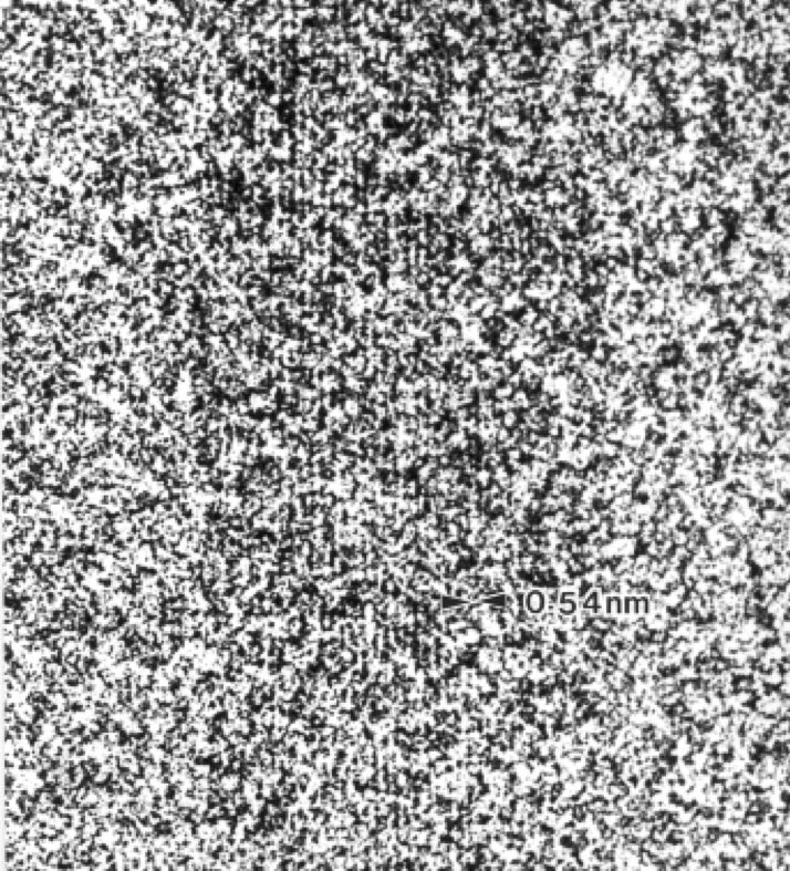
Direct visualization of cellulose lattice by electron microscopy (Master & Doctor study at Kyoto University)
I started my master study at KU in 1984 and worked on the above titled project under the supervision of the late Professor Harada. Scrutinized condition for low-dose high-resolution electron microscopy was realized for the first time to visualize the molecular arrangement of cellulose chains in a microfibril. The study ruled out the existence of crystalline subunit in a microfibril and demonstrated that the size and the state of crystallinity is species specific as originally proposed by Preston in early 1950s. The technique was applied to other natural and synthetic materials and provided new aspect in the polymer studies. People says “Seeing is not always believing in high resolution microscopy, but I learned ” Seeing is powerful if there is a sound background theory. Special thanks to Dr. Fujiyoshi, and Prof. Uyeda at KUICR.
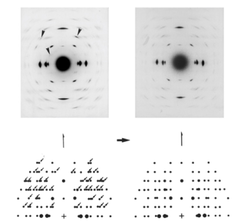
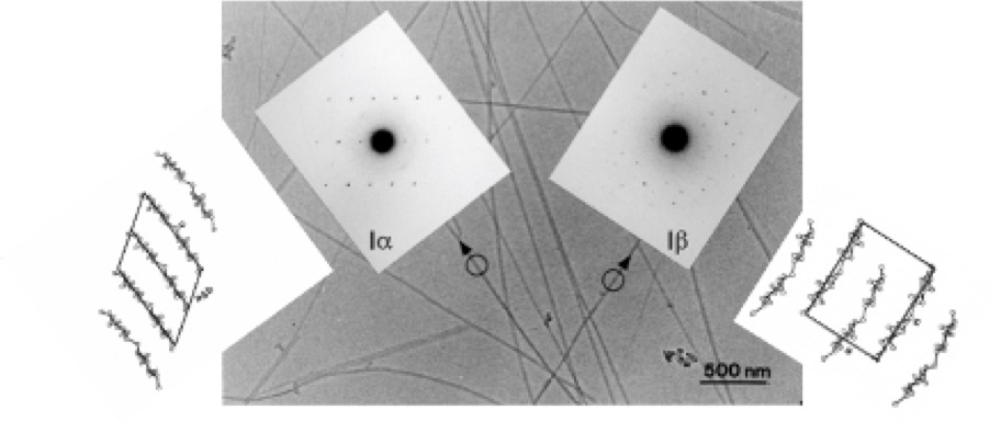
Crystal structure of cellulose Ialpha and Ibeta (Research Associate at The University of Tokyo, Post Doctoral fellow at CERMAV-CNRS)
I moved from KU to UT. Under Professor Okano’s direction, my interests start to shift from ‘seeing’ to ‘knowing’ the structure. Allomorphs of native cellulose crystal proposed by solid state NMR in 1980s has been long debated. The two-crystalline-phase concept was universally accepted when crystallographic evidence by electron micro-diffraction became available in 1991. The work was done at CERMAV-CNRS with Dr. Henri Chanzy. One-chain triclinic unit cell and two-chain monoclinic unit cell with different states of asymmetric units in crystals were determined, which lead to the proposal of the current crystal models of cellulose allomorphs. Localization of Ialpha and Ibeta in a microfibril was also proposed. Such crystallographic studies were also extended to other polymorphs such as cellulose II, IIII , beta chitin etc.
Biosynthesis and biodegradation of cellulose (Associate Professor at Wood Research Institute, Kyoto University)
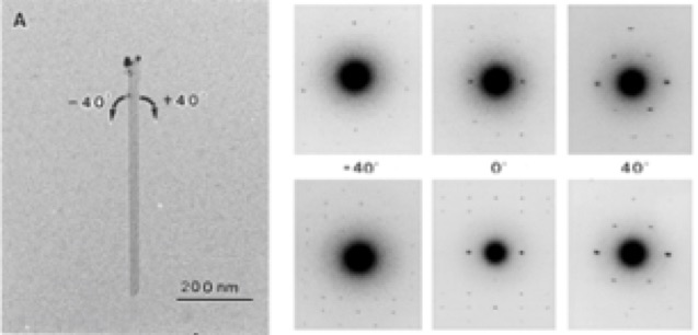
Back in KU but in Uji Research Campus. I start to analyse structure from the biological and biochemical point of view. This was a suggestion from one of my friend, a biochemist. Following his advice, I investigated how the cellulose molecules are polymerized, and how the cellulases deterioate micorofibril at the molecular level.
The molecular directionality of the chains in a unit cell was first determined experimentally by electron microdiffraction analysis of labeled microcrystals in 1997. This observation confirmed the “parallel-up” model of native cellulose. Having established the parallel-up model of cellulose, the directionality of the chains in a given microfibril could be identified simply by electron diffraction analysis. When nascent cellulose microfibril generated by Acetobacter xylinum was investigated, the reducing end of the growing cellulose chains was found to point away from the bacterium. This leads to the conclusion that the polymerization by cellulose synthase takes place at the non-reducing end of the growing cellulose chains. Following this successful application of electron diffraction and cytochemical staining reaction, biogenesis of cellulose and chitin and biodegradation by cellulases and chitinases were investigated in collaboration with domestic and oversea experts.
Cellulose nano-whiskers as a reinforced material (Associate Professor at Wood Research Institute, Kyoto University)
Also I involved in material science a little bit. Emerging interest of cellulose whiskers for the reinforcement filler for bulky polymers became interested during the second visit to CERMAV at France. It was early 1990s, under supervision of Dr Chanzy and Dr Maret, we demonstrated cellulose whiskers could be oriented when the non-flocculating suspension of cellulose microcrystals are placed in high static magnetic fields. Origins of this behavior, diamagnetic property, was examined by Cotton-Mouton effect and published recently (thanks to Bruno). After this experience, he was involved in several experiments on “biomimetic approach for making novel biomaterials”. I also contributed to fabricate transparent films made of nano-cellulose in its early stage of the development.
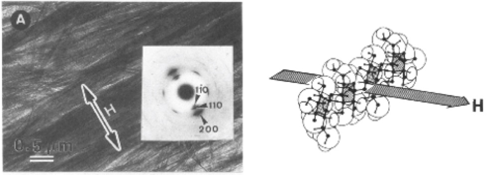
From Sugiyama J, Chanzy H, Maret G, The orientation of cellulose microcrystals by magnetic fields, Macromolecules, 25, 4232-4234, 1992
Biophysics and function of cellulose (Associate Professor at Wood Research Institute, and at Research Institute for Sustainable Humanosphere, Kyoto University)
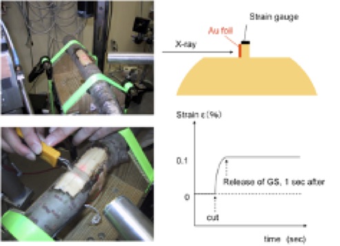
I was interested in the structure and properties relationship. To keep the branch at certain angle against the gravitational force, wood forms special tissue known as reaction wood. His primary interest was to know the role of cellulose in a tree. To keep the branch at certain angle against the gravitational force, wood forms special tissue known as reaction wood. His primary interest was to measure the state of cellulose whether it is under stress or not. Upper part of hardwood branch was placed precisely in the beam and the fiber repeat distance was measured before and after the release of the growth stress. This experiment for the first time evidenced that cellulose is under tension in tension wood. Related studies were carried out to learn structure – property relationship. Along this line, the project on going is: amazing tensile properties of cherry bark, accoustic properties of Picea wood in relation to their macro- to nano-structures.
Biomass analysis for sustainable utilization (Professor at Research Institute for Sustainable Humanosphere, Kyoto University)
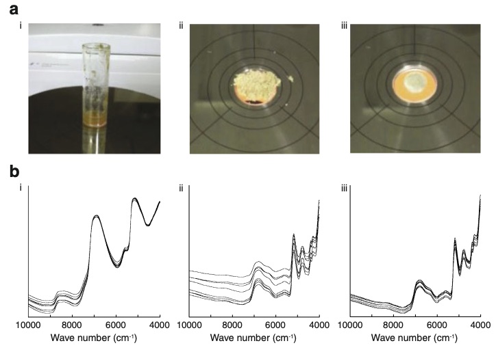
Basically, there are two subjects concerned. One is to develop a tool for rapid analysis to know the state of biomass before and after pretreatment and saccharification, for instance. The other is to visualize topochemical states of biomass during enzymatic processing to know the limiting factors that spoils the efficiency of saccharification.
Xyrarium related activity: Wood identification combined with machine learning (Professor at Research Institute for Sustainable Humanosphere, Kyoto University)
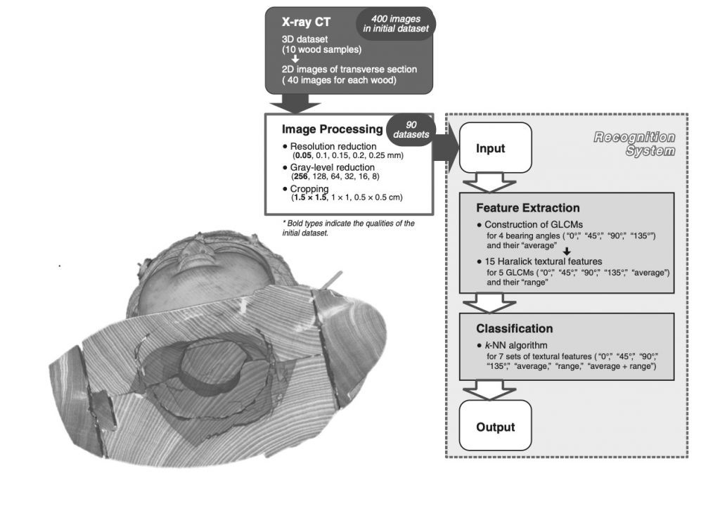
One of good examples of his activity is wood identification. To understand what wood species were used in the cultural artifacts is important in terms of their preservation and inheritance. However, a nondestructive method is required, and wood samples must be partly cut off in conventional methods such as microscopy. Therefore, he constructed a novel system for wood identification using image recognition of X-ray computed tomography images of major species used in Japanese wooden sculptures in collaboration with Kyushu National museum.
Representative papers
1) Direct visualization of cellulose lattice by electron microscopy
- Imai T, Putaux J-L, Sugiyama J , Geometric phase analysis of lattice images of crystalline allomorphs of Cladophora microcrystals, Polymer, 44, 1871-1879, 2003
- Helbert W, Nishiyama Y, Okano T, Sugiyama J, Molecular imaging of Halocynthia papillosa cellulose, J Struct Biol,124, 42-50, 1998
- Sugiyama J, Harada H, Saiki H, Crystalline morphology of Valonia cellulose IIII revealed by direct lattice imaging, Int J Biol Macromol, 9, 122-130, 1987
- Sugiyama J, Otsuka Y, Murase H, Harada H, Toward direct imaging of cellulose microfibrils in wood, Holzforschung, 40, 31-36, 1986
- Sugiyama J, Harada H, Fujiyoshi Y, Uyeda N , Lattice images from ultrathin sections of cellulose microfibrils in the cell wall of Valonia macrophysa Kutz ,Planta, 166, 161-168, 1985
- Sugiyama J, Harada H, Fujiyoshi Y, Uyeda N , Observations of cellulose microfibrils in Valonia macrophysa by high resolution electron microscopy, Mokuzai Gakkaishi , 31, 61-67, 1985
- Sugiyama J, Harada H, Fujiyoshi Y, Uyeda N, High resolution observations of cellulose microfibrils, Mokuzai Gakkaishi , 30, 98-99, 1984
2) Crystal structure of cellulose Ia and Ib
- Horikawa Y, Sugiyama J, Localization of Crystalline Allomorphs in Cellulose Microfibril, Biomacromolecules, 10, 2235-2239, 2009
- Mueller M, Burghammer, M, and Sugiyama J, Direct investigation of the structural properties of tension wood cellulose microfibrils using microbeam x-ray fibre diffraction, Holzforschung, 60, 474-479, 2006
- Kim N-H, Imai T, Wada M, Sugiyama J, Molecular directionality in cellulose polymorphs, Biomacromolecules, 7, 274-280, 2006
- Wada M, Heux L, Sugiyama J, Polymorphism of cellulose I family: reinvestigation of cellulose IVI, Biomacromolecules, 5, 1385-1391, 2004
- Šturcová A, His I, Apperley DC, Sugiyama J, Jarvis MC, Structural details of crystalline cellulose from higher plants, Biomacromolecules, 5, 1333-1339, 2004
- Hult E-L, Yamanaka S, Ishihara M, Sugiyama J , Aggregation of microfibrils in bacterial cellulose induced by high pressure incubation , Carbohydr Polym, 53, 9-14, 2003
- Hult E -L, Iversen T, Sugiyama J, Characterization of the supermolecular structure of cellulose in wood pulp fibres , Cellulose, 10, 103-110, 2003
- Nishiyama Y, Sugiyama J, Chanzy H, Langan P, The crystal structure and hydrogen bonding system in cellulose Ia, J Am Chem Soc, 125, 14300-14306, 2003
- Matthews JF, Skopec CE, Mason PE, Succato P, Torget RW, Sugiyama J, Himmel ME, Brady JW, Computer simulations of water structuring adjacent to microcrystalline cellulose Iβ structure, Carbohydr Res, 341, 138-152, 2006
- Hult E -L, Iversen T, Sugiyama J, Characterization of the supermolecular structure of cellulose in wood pulp fibres , Cellulose, 10, 103-110, 2003
- Tokoh C, Takabe K, Sugiyama J, Fujita M, CP/MAS13C NMR and electron diffraction study of bacterial cellulose structure affected by cell wall polysaccarides, Cellulose, 9, 351-360, 2002
- Tokoh C, Takabe K, Sugiyama J, Fujita M, Cellulose synthesized by Acetobacter xylinum in the presence of plant cell wall polysaccharides, Cellulose, 9, 65-74, 2002
- M Wada, L Heux, A Isogai, Y Nishiyama, H Chanzy, J Sugiyama, Improved structural data of cellulose IIII prepared in supercritical ammonia, Macromolecules 34 (5), 1237-1243, 2001
- Baker AA, Helbert W, Sugiyama J, Miles MJ, New insight into cellulose structure by atomic force microscopy shows the Ia crystal phase at near-atomic resolution, Biophys J, 79, 1139-1145, 2000
- Imai T, Sugiyama J, Itoh T, Horii F, Almost pure I cellulose in the cell wall of Glaucocystis, J Struct Biol, 127, 248-257, 1999
- Imai T, Sugiyama J, Nanodomains of Ia and Ib cellulose in algal microfibrils, Macromolecules, 31, 6275-6279, 1998 (104)
- A Baker, W Helbert, J Sugiyama, MJ Miles, High-Resolution Atomic Force Microscopy of NativeValoniaCellulose I Microcrystals, Journal of structural biology 119 (2), 129-138, 1997
- M Wada, T Okano, J Sugiyama, Synchrotron-radiated X-ray and neutron diffraction study of native cellulose Cellulose 4 (3), 221-232, 1997
- Wada M, Okano T, Sugiyama J, Horii F, Characterization of tension and normally lignified wood cellulose in Populus maximowiczii, Cellulose, 2, 223-233, 1995
- Heiner AP, Sugiyama J, Teleman O, Crystalline cellulose I and I studied by molecular dynamics simulation, Carbohydr Res, 273, 207-223, 1995
- Woodcock S, Henrissat B, Sugiyama J, Docking of congo red to the surface of crystalline cellulose using molecular mechanics, Biopolymer, 36, 201-210, 1995
- Wada M, Sugiyama J, Okano T, Two crystalline phase (Ia/Ib) system of native celluloses in relation to plant phylogenesis, Mokuzai Gakkaishi, 41, 186-192, 1995
- Wada M, Sugiyama J, Okano T, Native celluloses on the bases of two phase (Iα/Iβ) system J Appl Polym Sci, 49, 1491-1496, 1993
- Debzi EM, Chanzy H, Sugiyama J, Tekely P, Excoffier G, The Ia-b transformation of highly crystalline cellulose by annealing in organic solvents, Macromolecules, 24, 6816-6822, 1991
- Sugiyama J, Vuong R, Chanzy H, An electron diffraction study on the two crystalline phases occurring in native cellulose from algal cell wall, Macromolecules, 24, 4168-4175, 1991
- Sugiyama J, Persson J, Chanzy H, A combined infrared and electron diffraction study of the polymorphism of native celluloses, Macromolecules, 24, 2461-2466, 1991
- Sugiyama J, Okano T, Yamamoto H, Horii F, Transformation of Valonia cellulose crystals by an alkaline hydrothermal treatment, Macromolecules, 23, 3196-3198, 1991
3) Biosynthesis and biodegradation of cellulose
- Hult E-L, Katouno F, Uchiyama T, Watanabe T, Sugiyama J, Molecular directionality in crystalline β-chitin: hydrolysis by chitinases A and B from Serratia marcescens 2170, Biochem J, 388, 851-856, 2005
- Watanabe T, Ariga Y, Sato U, Toratani T, Nikaido N, Kezuka Y, Nonaka T, Sugiyama J, Aromatic residues within the substrate-binding cleft of Bacillus circulans chitinase A1 are essential for crystalline chitin hydrolysis, Biochem J, 376, 237-244, 2003 (84)
- Imai T, Watanabe T, Yui T, Sugiyama J, The directionality of chitin biosynthesis – A revisit, Biochem J, 374, 755-760, 2003
- Lehtio J, Sugiyama J, Gustavsson M, Fransson L, Teeri TT, The binding specificity and affinity determinants of the family 1 cellulose-binding domains from Trichoderma reesei Cel6A and Cel7A , Proc Natl Acad Sci USA, 100, 484-489, 2003 (271)
- Imai T, Watanabe T, Yui T, Sugiyama J , Directional degradation of β-chitin by chitinase A1 revealed by a novel reducing end labelling technique, FEBS Lett, 510, 201-205, 2002
- Uchiyama T, Katouno F, Nikaidou N, Nonaka T, Sugiyama J, Watanabe T , Roles of the exposed aromatic residues in crystalline chitin hydrolysis by chitinase A from Serratia marcescens 2170 , J Biol Chem, 276, 41343-41349, 2001 (137)
- Hashimoto M, Ikegami T, Seino S, Ohuchi N, Fukada H, Sugiyama J, Shirakawa M, Watanabe T, Expression and characterization of the chitin-binding domain of chitinase A1 from Bacillus circulans WL-12, J Bacteriol , 182, 3045-3054, 2000 (110)
- Sugiyama J, Boisset C, Hashimoto M, Watanabe T, Molecular directionality of b-chitin biosynthesis, J Mol Biol , 286, 247-255, 1999
- Imai T, Boisset C, Igarashi K, Samejima M, Sugiyama J, Unidirectional processive action of cellobiohydrolase Cel7A on Valonia cellulose, FEBS Lett, 432, 113-116, 1998 (87)
- Samejima M, Sugiyama J, Igarashi K, Eriksson K-EL , Enzymatic hydrolysis of bacterial cellulose, Carbohydr Res , 305, 281-288, 1998
- Hayashi N, Sugiyama J, Okano T, Ishihara M , The enzymatic susceptibility of cellulose microfibrils of the algal-bacterial type and the cotton-ramie type , Carbohydr Res , 305, 261-269, 1997
- Hayashi N, Sugiyama J, Okano T, Ishihara M, Selective degradation of cellulose Iα component in Cladophora cellulose with Trichoderma viride cellulase, Carbohydr Res , 305, 109-116, 1997
- Koyama M, Helbert W, Imai T, Sugiyama J, Henrissat B, Parallel-up structure evidences the molecular directionality during biosynthesis of bacterial cellulose, Proc Natl Acad Sci USA, 94, 9091-9095, 1997 (227)
- Gilkes NR, Kilburn DG, Miller RC Jr, Warren RAJ, Sugiyama J, Chanzy H, Henrissat B, Visualization of the adsorption of a bacterial endo-β-1,4-glucanase and its isolated cellulose-binding domain to crystalline cellulose, Int J Biol Macromol , 15, 347-351, 1993
4) Cellulose nano-whiskers as a reinforcement material
- B Frka-Petesic, J Sugiyama, S Kimura, H Chanzy, G Maret, Negative Diamagnetic Anisotropy and Birefringence of Cellulose Nanocrystals, Macromolecules, 48, 24, 8844-8857, 2015
- Thi Thi Nge, Masaya Nogi, Hiroyuki Yano, Junji Sugiyama, Microstructure and mechanical properties of bacterial cellulose/chitosan porous scaffold, Cellulose, 17, 349-363, 2010
- Yokota S, Kitaoka T, Sugiyama J, Wariishi H, Cellulose I nanolayers designed by self-assembly of its thiosemicarbazone on a gold substrate, Adv Mater, 19, 3368-3370, 2007
- Kvien I, Sugiyama J, Votrubec M, Oksman K, Characterization of starch based nanocomposites, J Mater Sci, 42, 8163-8171, 2007
- Nge Thi Thi, and Sugiyama J, Surface functional groups dependent apatite formation on bacterial cellulose microfibrils network in a simulated body fluid, J Biomed Mater Res, 81A, 124-134, 2006
- Uraki Y, Nemoto J, Otsuka H, Tamai Y, Sugiyama J, Kishimoto T, Ubukata M, Yabu H, Tanaka M, Shimomura M, Honeycomb-like architecture produced by living bacteria, Gluconacetobacter xylinus, Carbohydr Polym, 69, 1-6, 2007
- Nge Thi Thi, and Sugiyama J, Surface functional groups dependent apatite formation on bacterial cellulose microfibrils network in a simulated body fluid, J Biomed Mater Res, 81A, 124-134, 2006
- Yano H, Sugiyama J, Nakagaito AN, Nogi M, Matsuura T, Kurihara T, Handa K, Optical transparent composites reinforced with networks of bacterial nanofibers, Adv Mater, 17, 153-155, 2005
- Yamanaka S, Ishihara M, Sugiyama J, Structural modification of bacterial cellulose, Cellulose, 7, 213-225, 2000
- Matsumura H, Sugiyama J, Glasser WG , Cellulosic nanocomposites I Thermally deformable cellulose hexanoates from heterogeneous reaction, J Appl Polym Sci , 78, 2242-2253, 2000 (77)
- Ishikawa A, Sugiyama J, Okano T, Fine structure and tensile properties of ramie fibers in the crystalline form of cellulose I, II, IIII, and IVI, Polymer, 38, 463-468, 1997 (132)
- Sugiyama J, Chanzy H, Maret G, The orientation of cellulose microcrystals by magnetic fields, Macromolecules, 25, 4232-4234, 1992 (162)
5) Biophysics and function of cellulose
- Shengcheng Zhai, Yoshiki Horikawa, Tomoya Imai, and Junji Sugiyama, Cell wall characterization of windmill palm (Trachycarpus fortunei) fibers and its functional implications, IAWA Journal, 34, 20-33, 2013
- Bruno Clair, Tancrede Almeras, Gilles Pilate, Delphine Jullien, Junji Sugiyama, Christian Riekel, Maturation Stress Generation in Poplar Tension Wood Studied by Synchrotron Radiation Microdiffraction, Plant Physiology, 155, 562-570, 2010
- Wang Y, Gril J, Sugiyama J. Variation in xylem formation of Viburnum odoratissimum var. awabuki: growth strain and related anatomical features of branches exhibiting unusual eccentric growth , Tree Physiology, 29, 707-713, 2009
- Clair B, Gril J, Baba K, Thibaut B, Sugiyama J, Precautions for the structural analysis of the gelatinous layer in tension wood, IAWA J, 26, 189-195, 2005
- Clair B, Almeras T, Yamamoto H, Okuyama T, Sugiyama J, Mechanical state of native cellulose microfibrils in tension wood, Biophys J, 91, 1128-1135, 2006
- Kamiyama T, Suzuki H, Sugiyama J, Studies of the structural change during deformation in Cryptomeria japonica by time-resolved synchrotron small-angle X-ray scattering, J Struct Biol, 151, 1-11, 2005
- Clair B, Thibaut B, Sugiyama J, On the detachment of gelatinous layer in tension wood fibre, J Wood Sci, 51, 218-221, 2005
- Hori R, Suzuki H, Kamiyama T, Sugiyama J, The variation of microfibril angles and chemical composition-implication for functional properties, J Materi Sci Lett, 22, 963-966, 2003
- Hori R, Sugiyama J, A combined FT-IR microsocopy and principal component analysis on softwood cell walls, Carbohydr Polym, 52, 449-453, 2003
- Hori R, Mueller M, Watanabe U, Lichitenegger H, Fratzl P, Sugiyama J, The importance of seasonal differences in the cellulose microfibril angle in softwoods in determining acoustic properties, J Materi Sci, 37, 4279-4284, 2002
6) Biomass analysis for sustainable utilization
- Horikawa, Y., Imai, M., Kanai, K., Imai, T., Watanabe, T., Takabe, K., Kobayashi Y., Sugiyama, J., Line monitoring by near-infrared chemometric technique for potential ethanol production from hydrothermally treated Eucalyptus globulus, Biochemical Engineering Journal, 97, 65-72, 2015
- Horikawa, Y., Konakahara, N., Imai, T., Kentaro, A., Kobayashi, Y., Sugiyama, J., The structural changes in crystalline cellulose and effects on enzymatic digestibility, Polymer Degradation and Stability, 98, 2351-2356, 2013
- Horikawa, Y., Imai, T., Takada, R., Watanabe, T., Takabe, K., Kobayashi, Y., Sugiyama, J, Chemometric analysis with near-infrared spectroscopy for chemically pretreated Erianthus toward efficient bioethanol production, Applied Biochemistry and Biotechnology, 166, 711-721, 2012
- Horikawa, Y., Imai, T., Takada R., Watanabe, T., Takabe, K., Kobayashi Y., Sugiyama, J., Near-Infrared Chemometric Approach to Exhaustive Analysis of Rice Straw Pretreated for Bioethanol Conversion, Applied Biochemistry and Biotechnology, 164, 194-203, 2011
7) Xylarium related activity
- Hwang SW, Lee WH, Horikawa Y, Sugiyama J, Identification of Pinus species related to historic architecture in Korea by using NIR chemometric approaches, Journal of Wood Science, 62(1), 156-167, 2016/02
- Hwang SW, Lee WH, Horikawa Y, Sugiyama J, Chemometrics approach for species identification of Pinus densiflora Sieb. et Zucc. and Pinus densiflora for. erecta Uyeki (in Korean), J Kor Wood Sci Technol, 43, 6, 701-713, 2015
- Kobayashi Kayoko, Akada Masanori, Torigoe Toshiyuki, Imazu Setsuo, Sugiyama Junji, Automated recognition of wood used in traditional Japanese sculptures by texture analysis of their low-resolution computed tomography data, Journal of Wood Science, 61(6), 630-640, 2015/09
- Rie Endo, Tsutomu Hattori, Makoto Tomii, Junji Sugiyama, Identification and conservation of a Neolithic polypore, Journal of Cultural Heritage, 16, 869-875,2015/05
- Ugai Watanabe, Hisashi Abe, Kazumasa Yoshida, Junji Sugiyama, Quantitative evaluation of properties of residual DNA in Cryptomeria japonica wood, Journal of Wood Science, 61(1), 1-9, 2015/01
- Horikawa, Y., Mizuno-Tazuru, S., Sugiyama, J., Near-infrared spectroscopy as a potential method for identification of anatomically similar Japanese diploxylons, Journal of Wood Science, 61(3), 251-261, 2015
- Mertz, M., Gupta, S., Hirako, Y., Azevedo P., Sugiyama, J., Wood selection of ancient temples in the Sikkim Himalayas, IAWA J, 35(4), 444-462, 2014
- Yumiko Watanabe, Shigeki Tamura, Takeshi Nakatsuka, Suyako Tazuru, Junji Sugiyama, Bambang Subiyanto, Toshitaka Tsuda, and Takahiro Tagami, Comparison of Sungkai Tree-Ring Components and Meteorological Data from Western Java, Indonesia, Journal of Disaster Research , 18, 95-03, 2013
- Mizuno, S, Torizu, R. Sugiyama, J, Wood identification of a wooden mask using synchrotron X-ray microtomography, J. Archeological Sci., 37, 2842-2845, 2010
